 Home
Home
|
Table of Content - Volume 19 Issue 1 - July 2021
Computed tomography in diagnosis of acute appendicitis
Pratap M Jadhav1*, Vaishali S Choure2, Ojal Pratap Jadhav3
1Professor, Department of Radiology, Al-Azar Medical College, Ezhalloor, Thodupuza, Kerala, INDIA. 2Consultant, Department of Anesthesiology, SNDH and Research Centre, Aurangabad, Maharashtra, INDIA. 3MBBS. Aurangabad, INDIA. Email: drjadhavpm@gmail.com
Abstract Background: The study aimed to assess the diagnostic accuracy of computed tomography (CT) in diagnosing acute appendicitis in clinically suspected patients at a tertiary superspeciality hospital in rural area. Method: A total 190 patients (102 male and 88 female; average range 15-70 years) with clinically suspected acute appendicitis underwent non-enhanced helical CT, over a period of one year. Criteria for diagnosis of acute appendicitis included appendiceal size (> 6mm diameter) and presence of other signs of appendicitis. CT results were compared with surgical and pathological findings. Result: Based on the results of studies, sensitivity and specificity of CT in the diagnosis of acute appendicitis were 97.32% and 88.42% respectively. An overall accuracy was 96.8%. Conclusion: CT is fast, cost-effective and an accurate modality in diagnosing acute appendicitis in clinically suspected patients. Key words: Acute appendicitis, Computed tomography (CT)
INTRODUCTION Acute appendicitis is one of the most common abdominal emergencies1,2. It’s progression to perforation is associated with high morbidity and mortality rates2. There has been continuous search for complementary diagnostic method to limit the number of “unnecessary” appendectomies without delaying the diagnostic and therapeutic process and without increasing perforation rate. The Alvarado scoring system is used for the diagnosis of acute appendicitis, and it is based mainly on patient history, clinical examination and simple blood tests2,3,4. In his original paper, Alvarado5 recommends surgery for patients with score equal to or greater than 7. However, the use of imaging modalities such as ultrasonography (US) and computed tomography (CT) has helped to decrease the morbidity and mortality1,6. In the present study, CT was performed to assess the diagnostic accuracy in diagnosing acute appendicitis in clinically suspected patients.
METHODS Over a one-year period, 190 patients (102 male and 88 female) with clinically suspected acute appendicitis underwent non-enhanced helical CT. Inclusion criteria for clinically equivocal acute appendicitis were based on the clinical judgement of the referring surgeon. All CT scans were obtained with a helical CT scanner (HiSpeed Advantage; GE Medical system). Axial images were obtained from L2 vertebra level to symphysis pubis with a slice thickness of 5-mm. All scans were obtained without oral, rectal and intravenous contrast. CT scan findings were defined as presence of at least one of the following parameters: 1) outer to outer diameter > 6mm; 2) peri-appendiceal fat stranding; 3) appendicolith; 4) peri-appendiceal free fluid; 5) phlegmon; and 6) abscess formation1,6,7. The CT findings were compared with the surgical and histopathological reports. All patients who did not undergo surgery were followed up clinically.
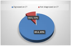
RESULTS All 190 patients included were subjected to CT scan within 2-4 hours of presentation and underwent operative intervention. Amongst the patients included in the study, 170 patients (89.4%) had CT scan findings suggestive of acute appendicitis and 20 (10.5%) did not meet the CT scan criteria of acute appendicitis. The most common finding on CT scan was peri-appendiceal fat stranding in 124 patients (65.3%). Other findings were outer to outer diameter more than 6mm in 60 patients (31.6%), presence of appendicolith in 40 patients (21.1%) and peri-appendiceal fluid in 22 patients (11.6%). Less frequent findings were presence of phlegmon formation in 8 patients (4.2%) and abscess formation in 4 patients (2.1%). Amongst the 170 patients (89.4%) who were found to have CT scan findings suggestive of acute appendicitis, 168 patients (88.4%) were confirmed to have acute appendicitis intra-operatively. Additional, of the 20 patients (10.5%) who did not demonstrate the findings on CT scan, 4 patients (2.1%) were confirmed to have acute appendicitis intra-operatively. Overall, the study demonstrated that in suspected acute appendicitis, a non-enhanced CT scan is 97.32% sensitive, 88.42% specific, 96.8% accurate with a positive predictive value of 98.8% and negative predictive value of 80.0%.
Table 1: CT Findings in patients suggestive of Acute appendicitis
Table 2: Statistical Analysis
Figure 1: Showing Frequency of CT scan based diagnosis of Acute appendicitis.
DISCUSSION An accurate diagnosis of acute abdomen is important in distinguishing surgical conditions like acute appendicitis from nonsurgical conditions that may have a similar presentation. Various pathologies like right sided diverticulitis, cecal carcinoma, mesenteric inflammation, crohn’s colitis, ectopic pregnancy and complicated ovarian cysts may mimic on CT imaging8. A surgeon’s clinical evaluation alone can reliably diagnose acute appendicitis without help of CT. Nevertheless, the surgeon’s assessment may miss the cases in patients meeting few diagnostic criteria. It is these patients in whom CT becomes and effective and useful adjunct in the workup of acute appendicitis. In the literature the rate of negative appendectomy has been reported as high as 17-36% without the use of CT9. A recent meta-analysis suggested that CT in suspected appendicitis patients could reduce the rate of unnecessary surgery without increasing morbidity10. In summary, CT in patients with suspected acute appendicitis can be extremely helpful not only to diagnose acute appendicitis but also to establish an alternate diagnosis. In our study CT scan is 97.32% sensitive, 88,42% specific.
REFERENCE
Policy for Articles with Open Access
|
|
 This work is licensed under a Creative Commons Attribution-NonCommercial 4.0 International License.